Dog Eye Problems
What do dogs see?
2017 Study finds high visual acuity revealed in dogs (pdf)Lind O, Milton I, Andersson E, Jensen P, Roth LSV (2017) High visual acuity revealed in dogs. PLOS ONE 12(12): e0188557.
Quick Facts
- Dogs see much better in the dark
- Dogs see motion and quick movement better than humans
- Dogs to not see colors like humans do
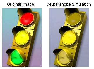
- Dogs are unable to distinguish between green, yellow, orange and red (imagine a dog driving and watching a traffic light)
- Dogs have a hard time differentiating between greens and grays
- Dogs have less binocular vision than humans and have a larger field of view
- Dogs don't see approsimately 6 times less detail than humans
http://www.vischeck.com/
http://www.diycalculator.com/sp-cvision.shtml#A8b

Hunting retrievers use all of their senses while working - and we depend heavily on their use of the sense of sight in performing their role. However, our visual perception of the world is different than our dogs, and a better understanding of the differences in our visual abilities may help us better understand how our hunting dogs work. Recently, an article was published in the veterinary literature that reviewed our current understanding of vision in normal dogs. This article, titled
"Vision in dogs", was written by Paul E. Miller, DVM and Christopher J. Murphy, DVM, and published in the Journal of the Veterinary Medical Association (JAVMA, vol. 207, no. 12, pp. 1623-1634, Dec. 15, 1995). The purpose of this article is to discuss the information provided by Drs. Miller and Murphy, and occasionally interject my interpretation of how this material pertains to our hunting retrievers. Please bear in mind that the original authors deserve the credit for compiling the
following information! I would encourage anyone that wishes to get the "whole story" to obtain a copy of the complete article.
Fundamentals of vision
A number of factors are involved in vision; it includes not only the perception of light and motion, but also visual perspective, visual field of view, depth perception, visual acuity, and the perception of color and form. Our ability to describe these factors is based on our own perception, and we may not be able to accurately describe canine vision because of fundamental differences in vision between the two species, and also because of our understanding of some of these factors in the canine
is imperfect.
Sensitivity to light
The authors start their discussion of the canine visual system with a fundamental difference between canine and human vision. The canine visual system is designed to operate well under low light conditions, while the human visual system performs best in bright light. The canine visual system is capable of functioning under a wide range of lighting conditions, but the adaptation to low light conditions enhance their ability to function as predators. The minimum threshold of light for vision in
cats is six times lower than that for humans, and the although the minimum threshold for dogs is thought to somewhat greater than that for cats, it is still much lower than that for humans.
Several methods are used by dogs to improve vision in low light conditions. The principal method is based on the types of photoreceptors present in the canine retina. The retina of the dog's eye is composed primarily of rod photoreceptors, while the human retina is composed primarily of cone photoreceptors. Rods function better in dim light, while cones are used for color vision and require bright light. Humans and canines both have rods and cones present in their retina's, but the
relative amount of each is very different.
Another method used by dogs to improve low light vision is the use of a tapetum lucidum. The tapetum lucidum is a highly reflective layer of cells located behind the photoreceptors in the canine retina. This reflective layer is responsible for the bright shine of a dog's (and other species) eye when a bright light is shone at the eye in the dark. This reflective layer functions to improve low light vision by reflecting light back through the retina, essentially allowing the photoreceptors
two chances to react to each quantum of light. The tapetum lucidum may also function in another manner. In other species, it has been shown that the tapetum lucidum shifts the wavelength (via fluorescence) of the reflected light to more closely match the optimal wavelength for the sensitivity of the rod photoreceptors, thus enhancing contrast.
The tapetum lucidum is located in the top half of the dog's retina (termed the tapetal area of the retina); the bottom half of the dog's retina is composed of the tapetum nigrum, a layer of darkly pigmented cells that is not reflective. In most instances, the top half of the retina receives light from the darker ground, and the bottom half of the retina receives light form the brighter sky. This is felt to enhance the view of both the darker ground and brighter sky.
Sensitivity to motion
Little work has been done on this subject, although rod photoreceptors are better able to detect motion and shapes than cones. An early study indicated that dogs could discriminate an object in motion at 810 to 900 meters, but were only able to discriminate the same object when stationary at 585 meters or less.
Sensitivity to flickering lights
The frequency at which rapidly flickering light fuses appears to fuse into a constantly illuminated light is termed "flicker fusion" and can be used by investigators to gain information about the retinal rods and cones. In most humans, flicker fusion occurs at around 50 to 60 Hz, although some people can detect flicker up to about 70 Hz. It appears that the flicker fusion rate for most dogs may be as high as 70 to 80 Hz. How is this important to hunting retrievers? This may explain
why our dogs don't spend much time watching television! The refresh rate on televisions is about 60 Hz, hence we perceive the television image as a smooth image, but to our dogs it may appear as a rapidly flickering image.
Visual perspective
The height of the eyes above the ground has a major effect on the view that an animal or person sees. This is readily apparent if one crouches down and watches a mark from your dog's eye level, and the importance of evaluating marks from this perspective when setting up training scenarios or hunt tests is well known.
Visual field of view
Visual field of view is the area seen by an eye when it is fixed on one point. In dogs, this has not been well studied, and probably varies between breeds, because of the differences in the shape of the skull, placement of the eyes in the skull, and the shape and size of the nose. In the average dog, the eyes are placed such that they deviate approximately 20 degrees lateral to the midline. In humans, the eyes do not deviate, but rather look straight ahead. When both eyes are considered
together, two studies have indicated that the total field of view in dogs is approximately 240 to 250 degrees, which is 60 to 70 degrees greater than the normal human's field of view (180 degrees). Our dogs are probably more aware of activity occurring around them than we are because of this larger field of view.
Depth perception
Our ability to determine depth depends principally upon binocular vision, which is present in the region of the field of view where the visual fields of each eye overlap. Various studies have resulted in several different estimates of the degree of binocular vision present in dogs, and this again may vary between breeds because of anatomical differences in skull shape. It appears that the area of visual overlap is approximately 30 to 60 degrees in most dogs, which is much less than in humans,
who have approximately 140 degrees of binocular overlap.
Monocular depth perception is also possible, however. A number of clues can be used to indicate depth, such as relative brightness, contour, areas of light and shadow, object overlay, linear and aerial perspective, and density of optical texture. Movement of the head can also be used to assist in depth perception. Although the area of binocular depth perception available to our dogs is smaller than that available to us, it is obvious to all of us with hunting retrievers that they can clearly
judge distances very well.
Visual acuity
Visual acuity is the ability to see the details of an object separately and clearly. Visual acuity depends on three factors: a) optical properties of the eye, b)retinal detection and processing of the image, and c) proper interpretation of the images by higher centers in the brain. Postretinal processing has not been extensively studied in dogs, and was beyond the scope of the article being reviewed.
Optical factors in visual acuity:
The optical media of the eye consists of the cornea, aqueous humor, lens, and vitreous humor. These structures are responsible for creating a properly focused image on the retina (emmetropia). If the image is focused in front of the retina, myopia, or nearsightedness, results, and if the image is focused behind the retina, hyperopia, or farsightedness, results. Some dogs are myopic, or nearsighted. One study demonstrated that 53% of a group of German Shepherds were myopic, and another study
indicated that 64% of a group of Rottweilers were myopic. However, another study of a group of guide dog German Shepherds found only 15% of this group of dogs were myopic, indicating that selection for normal vision may have occurred. Myopic dogs may have been removed from the program due to poorer performance, without an understanding of why they were performing poorly. This finding may have important implications for retriever breeds, indicating that poor marking skills may possibly be
related to vision disturbances, and use of animals with poor marking skills in breeding programs may possibly propagate those vision problems. Other optical aberrations may occur within the eye, creating vision problems, such as astigmatism, spherical aberrations of the lens, and chromatic aberrations. The clinical importance of these conditions is unclear in dogs - they are either an uncommon finding or the canine eye may be able to accommodate for these problems.
In addition, the normal eye is able to accommodate, or change focus, which allows normal vision of objects at different distances. The canine eye has limited accommodative ability compared to the human eye. They appear to be only able to accurately image objects within 50 to 33 cm of their eye, where as human children are able to accurately image objects as close as 7 cm. Dogs compensate for this limited accommodative ability by using other senses, such as smell or taste, to augment vision of
very close objects. As humans age, it is normal to lose some accommodating ability (age-related presbyopia), and it is thought that dogs also undergo similar age related changes in accommodation, but the incidence or significance has not be studied in dogs.
Retinal factors in visual acuity:
It is felt that the retina is the principle limiting factor of visual acuity in dogs. To improve vision in dim light, a greater number of photoreceptors converge on a single ganglion cell (a nerve cell that gathers input from receptor cells and then transmits the information to higher nervous centers in the brain). The more photoreceptors converging on a single ganglion cell, the less detail is present in the image produced, just as high speed photographic film, designed for low light
situations, produces a grainier image than lower speed (brighter light) photographic film. The more ganglion cells present, the more nerve fibers present in the optic nerve that relays visual information to the brain. The canine optic nerve contains approximately 167,000 nerve fibers, compared with the human optic nerve which contains 1.2 million nerve fibers.
In the human eye, there is a circular area of the retina that contains the densest concentration of photoreceptors and ganglion cells, called the fovea. It is centrally located, and is the area of the retina that produces the sharpest image. The canine eye lacks a fovea, but rather has a region termed the visual streak. The visual streak is an oval area of the retina located just above the optic nerve, and is positioned with its long axis on a horizontal plane, and is centered on the area of
the retina closer to the nose. Like the fovea, it contains the highest concentration of photoreceptors and ganglion cells, and is the area that produces the greatest visual acuity. It is located in the tapetal area of the retina, which again aids in enhancing vision in dim light. The oval shape of the visual streak probably helps improve a dog's ability to scan the horizon, and the nasal location of the visual streak probably enhances a dog's ability to use peripheral vision.
Individual variations in the density of ganglion cells within the visual streak have been noted. Wolves have a higher density of ganglion cells than dogs (12,000 to 14,000 ganglion cells/mm2 in wolves compared to 6,400 to 14,400 ganglion cells/mm2 in dogs) which suggests that wolves have better visual acuity than dogs. Differences are found between dogs, also, and this may be another area where selection for visual ability may occur. One study indicated that one strain of Beagles had a
pronounced visual streak, whereas another strain had only a moderately pronounced streak. It is possible, although difficult, to differentiate between pronounced and moderate visual streaks with ophthalmoscopic evaluation, and this may potentially be a tool that could be used in selection of breeding stock. Careful evaluation of the retina by a competent ophthalmologist, using sophisticated equipment, is required to make this differentiation. Studies to validate the effectiveness of this
technique are required before this technique can be recommended in breeding stock selection.
Estimates of visual acuity:
The most commonly used indicator of visual acuity is the Snellen fraction, which relates the ability to distinguish objects or letters at a fixed distance with the standard response. Snellen fractions of 20/20, 20/40, 20/60 or 20/100 indicates that the test subject needs to be 20 feet away from an image to clearly visualize the details that a normal subject could discern at 20, 40, 60, and 100 feet away, respectively. A variety of studies have been done, using a number of different
methodologies, to estimate the visual acuity of dogs. We can assume from these studies that the normal dog has a visual acuity of approximately 20/75. This means that a dog must be 20 feet away from an object to clearly visualize details of that object that a human with normal vision could clearly visualize from a distance of 75 feet. Again, this less acute vision in dogs is a trade-off for improved vision in dim light, and their life style does not require visual distinction of fine details as
does our life style.
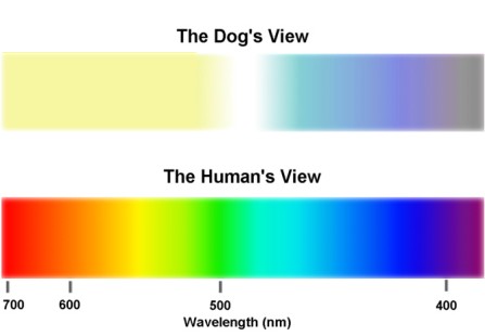
Color vision
A number of studies have been done to investigate the color vision of dogs, and the results have been conflicting. However, more recent, better controlled studies indicate that dogs do possess and use color vision, but not to the same degree that humans do. The photoreceptor used for color vision is the cone, and there are cones present in the canine retina. However, they are present in low numbers, comprising less than 10% of the total photoreceptor population in the central area of the
retina, as opposed to the human retina which consists of nearly 100% cones in the fovea. Two distinct type of cones appear to be present in the canine retina. One type is maximally sensitive to light in the wavelength that appears violet to people, and the other type is maximally sensitive to light in the wavelength that appears yellow-green to people. Thus, it appears that the visual spectrum of color in dogs is divided into two hues; one in the violet and blue-violet range, probably appearing
as blue, and the other in the greenish-yellow, yellow, and red range, which is probably seen as yellow. Light that appears blue-green to people probably appears as white or shades of gray to dogs. Dogs are unable to differentiate colors that appear as green, yellow-green, orange or red to people, and are unable to differentiate greenish-blue from gray. This is similar to people who are red-green color blind. However, one study indicates that dogs are better able to differentiate between subtle
shades of gray than people, which would be advantageous in increasing visual discrimination in low light conditions, where insufficient light is available to stimulate cones.
Summary
The authors conclude by stating that although the canine visual system may be considered inferior to the human visual system in such aspects as degree of binocular overlap, color perception, accommodative range, and visual acuity, the canine visual system is superior to the human visual system in other aspects, such as functional ability in low light conditions, retinal response rate to another image (flicker fusion), field of view, ability to differentiate shades of gray, and possibly the
ability to detect motion. The canine visual system is optimized to exploit a different environmental niche than our own, and hopefully by better understanding the strengths and weaknesses of the canine visual system we will be better able to understand our hunting companion's capabilities. Let me conclude by encouraging those of you interested in this issue to read the complete article, which includes 68 references to other works. I wish to personally thank the authors for greatly improving
my own understanding of the canine visual system.
http://psychlops.psy.uconn.edu/eric/class/dogvision.html
----------------------------------------------------------------------------------------
Owners who want to better understand their canine companions must recognize that dogs see the world from a different visual perspective. The differences begin with the structure of
the eye. "We have a good idea what canines see because we know the make-up of the retina of a dog's eye," says Dr. Ralph Hamor, a veterinarian and specialist in ophthalmology
at the University of Illinois College of Veterinary Medicine Teaching Hospital.
The retina, which covers the back of the inside of the eyeball, contains cones and rods-two types of light-sensitive cells. Cones provide color perception and detailed sight, while rods
detect motion and vision in dim light. Dogs, which have rod-dominated retinas, see better in the dark than humans do and have motion-oriented vision. However, because they have
only about one-tenth the concentration of cones that humans have, dogs do not see colors as humans do.
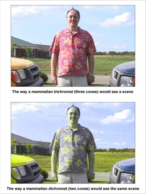
"I generally explain that dogs see like a color-blind human," says Dr. Hamor. "Many people think that a person who is red/green color blind cannot see any color, but there are
variations of being color blind. Most people have vision that is trichromatic (three color variations). People who are red/green color blind are dichromatic (two color variations).
Dogs can pick out two colors-blue-violet and yellow-and they can differentiate among shades of gray." Dogs are unable to distinguish among green, yellow, orange, and red. They
also have difficulty differentiating greens and grays.
Dogs use other cues (such as smell, texture, brightness, and position) rather than rely on color. Seeing-eye dogs, for example, may not distinguish whether a stoplight is green or
red; they look at the brightness and position of the light. This and the flow and noise of traffic will tell the dog that it is the right time to cross the street.
The set of dog's eyes determines the amount of field of view and depth perception. Prey species tend to have eyes set on the sides of their head because the increased field of view
allows them to see approaching predators. Predator species, like humans and dogs, have eyes set closer together. "Human eyes are set straight forward while dog eyes, depending
on the breed, are usually set at a 20 degree angle. This angle increases the field of view and therefore the peripheral vision of the dog."
However, this increased peripheral vision compromises the amount of binocular vision. Where the field of view of each eye overlaps, we have binocular vision, which gives us
depth perception. The wider-set eyes of dogs have less overlap and less binocular vision. Dogs' depth perception is best when they look straight ahead, but is blocked by their noses
at certain angles. "Predators need binocular vision as a survival tool," Dr. Hamor says. Binocular vision aids in jumping, leaping, catching, and many other activities fundamental to
predators.
In addition to having less binocular vision than humans, dogs also have less visual acuity. Humans with perfect eyesight are said to have 20/20 vision-we can distinguish letters or
objects at a distance of 20 feet. Dogs typically have 20/75 vision-they must be 20 feet from an object to see it as well as a human standing 75 feet away. Certain breeds have better
acuity. Labradors, commonly used as seeing-eye dogs, have been bred for better eyesight and may have closer to 20/20 vision.
Don't expect your dog to recognize you across the field by sight. He'll recognize you when you do some sort of motion particular to yourself or by smell or hearing. Because of the
number of rods in the retina, dogs see moving objects much better than they do stationary objects. Motion sensitivity has been noted as the critical aspect of canine vision. "So much
of dog behavior deals with posture and appropriateness. Small changes in your body posture mean a lot to your dog," Dr. Hamor adds. Dog owners need to modify training
based on this fact. If you want your dog to perform an action based on a silent cue from you, Dr. Hamor suggests using a wide sweeping motion to cue your dog.
When dogs go blind, owners often wonder if the dogs' quality of life has diminished to the point where they are no longer happy. "We know that humans deal well with being blind,
and humans are much more dependent on their eyes than are dogs," Dr. Hamor says. "Blind dogs lead happy lives if they are comfortable." The owner may need to make some
adjustments in the pet's environment, such as having a fenced yard, taking leashed walks, and not leaving unusual objects in normal pathways. "When blind dogs are in their normal
environment, most people don't know they are blind." When clients visit Dr. Hamor asking about quality of life for their newly blind dog, Dr. Hamor suggests that they take a month to
see if they and their dog are happy. In the majority of cases, the owners never come back.
http://vetmed.illinois.edu/petcolumns/showarticle.cfm?id=116